
Understanding and Treating Baby Skull Shape Abnormalities: Plagiocephaly, Craniosynostosis, and Beyond
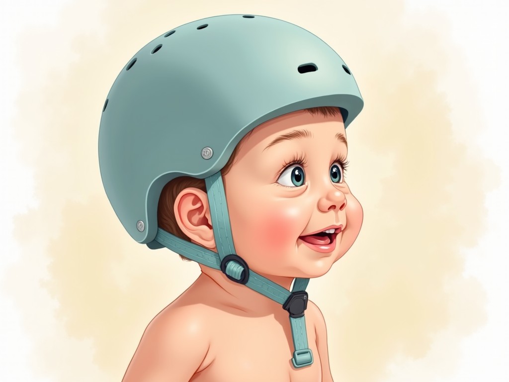
When a newborn’s head looks oddly shaped, parents often feel a mix of worry and curiosity. While many skull shape changes are harmless and resolve on their own, some require early intervention to prevent long‑term complications. This guide explains the most common skull shape abnormalities in infants, how they are diagnosed, and the evidence‑based treatment options available today.
What Are Baby Skull Shape Abnormalities?
Infant skulls are made of flexible, overlapping bones that grow rapidly during the first year of life. Because the bones are still soft, external pressure can influence the shape of the skull. Two main categories of skull shape problems exist:
- Positional Plagiocephaly – a flattening of the back or side of the head caused by prolonged pressure.
- Craniosynostosis – premature fusion of one or more skull sutures, restricting growth in that area.
In addition, certain genetic syndromes (e.g., Apert, Crouzon, Pfeiffer, Saethre‑Chotzen) can present with skull shape changes along with other facial and limb abnormalities.
Recognizing the Signs: When to Seek a Professional Evaluation
Parents should be alert to the following indicators:
- Visible flattening on the back or side of the head that does not improve with repositioning.
- Uneven hair distribution or a noticeably low hairline.
- Head turning consistently to one side (torticollis) or a stiff neck.
- Facial asymmetry, such as a sunken eye socket or a flattened nasal bridge.
- Any associated developmental delays or seizures.
Early detection is key. A pediatrician will take a detailed medical history and perform a physical exam. In many centers, a 3‑D scanner is used to capture precise skull measurements, aiding in diagnosis and treatment planning.
Diagnostic Pathways
Positional Plagiocephaly
Diagnosis is usually clinical, based on the pattern of flattening and the infant’s sleep position. Imaging is rarely required unless the shape is severe or atypical.
Craniosynostosis
Computed Tomography (CT) scans are the gold standard for confirming premature suture fusion. The scan shows which sutures are fused and the extent of skull deformity.
Genetic Syndromes
When skull shape abnormalities are accompanied by other anomalies, genetic testing and a multidisciplinary evaluation (including genetics, neurology, and craniofacial surgery) are recommended.
Treatment Options: From Gentle Measures to Surgery
Positional Plagiocephaly
Early intervention yields the best outcomes. The primary strategy is to reduce pressure on the affected area:
- Repositioning – during awake times, place the baby on the side or belly to relieve pressure.
- Physical Therapy – for infants with torticollis, stretching exercises help correct neck stiffness.
- Helmet Therapy – custom-fitted helmets distribute gentle pressure to guide skull growth. Helmets are most effective when started before 6 months of age.
- Monitoring – regular follow‑ups to assess progress and adjust treatment.
Craniosynostosis
Once a CT confirms suture fusion, surgical intervention is usually indicated to relieve intracranial pressure and allow normal brain growth:
- Open Craniotomy – the surgeon removes the fused suture and reshapes the skull. This is typically performed before 12 months of age.
- Endoscopic Strip Craniectomy – a minimally invasive option for selected cases, often combined with postoperative helmet therapy.
- Multistage Reconstruction – for complex or syndromic craniosynostosis, multiple surgeries may be required as the child grows.
Genetic Syndromes
Treatment is tailored to the specific syndrome and may involve:
- Multiple cranial surgeries to correct skull and facial bone abnormalities.
- Orthodontic and dental interventions for mid‑facial hypoplasia.
- Rehabilitation for associated motor or cognitive delays.
Key Takeaways for Parents
- Most positional plagiocephaly cases improve with simple repositioning and, if needed, helmet therapy.
- Early detection of craniosynostosis is critical; surgery before 12 months offers the best neurodevelopmental outcomes.
- Infants with genetic syndromes require a coordinated care team to address multiple health issues.
- Regular follow‑ups with a pediatric craniofacial specialist ensure timely adjustments to treatment plans.
Frequently Asked Questions
1. How long does helmet therapy last?
Typically 6–12 months, depending on the severity of the deformity and the infant’s age at initiation.
2. Is surgery risky?
Like any surgery, there are risks, but pediatric cranial surgeons have a high success rate and low complication rates when performed in specialized centers.
3. Can I use a baby carrier to prevent flattening?
Yes, using a carrier that distributes weight evenly and allows the baby to change positions can reduce pressure on the skull.
Conclusion
Baby skull shape abnormalities are common but manageable with timely, evidence‑based care. Whether the issue is positional flattening or a more complex craniosynostosis, a multidisciplinary approach—combining early detection, gentle repositioning, helmet therapy, and, when necessary, surgical intervention—offers the best chance for healthy growth and development. If you notice any irregularities in your infant’s head shape, schedule an evaluation with your pediatrician or a craniofacial specialist to ensure the right treatment plan is put in place.
Considering treatment in Türkiye?
Get a free consultation and cost estimate from verified specialists.