Spinal Compression Fracture: Diagnosis, Symptoms, and Modern Treatment Options
Spinal compression fractures are a common yet often overlooked injury that can dramatically affect quality of life. Whether caused by a sudden fall, a car accident, or the gradual wear of osteoporosis, these fractures can lead to chronic pain, loss of height, and even paralysis if not addressed promptly. In this guide, we break down the key facts—symptoms, diagnostic tools, and both conservative and surgical treatment strategies—so you can recognize the signs early and seek the right care.
Understanding Spinal Compression Fractures
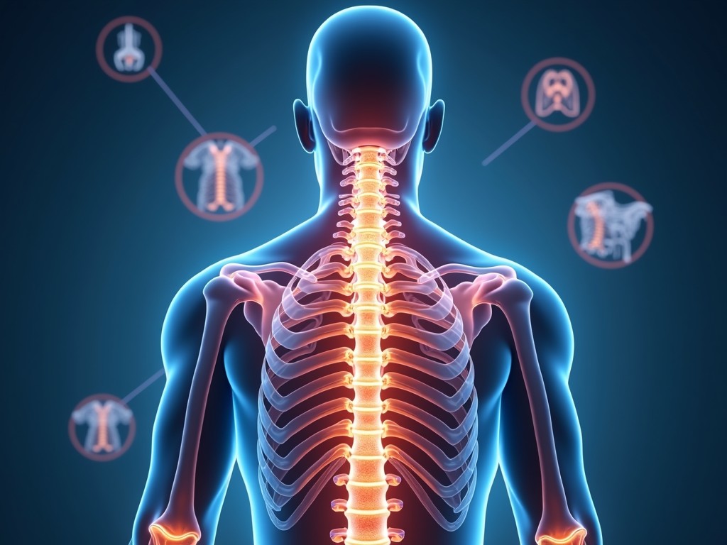
A spinal compression fracture occurs when one or more vertebrae collapse under pressure. The most frequent sites are the thoracic (mid-back) and lumbar (lower back) regions, especially the junction between the 12th thoracic and 1st lumbar vertebrae (T12–L1). While 70% of fractures happen in the thoracolumbar area, 5–10% affect the cervical spine, and the remainder involve the sacrum or coccyx.
Common Causes
- High‑impact trauma: car accidents, sports injuries, or falls from height.
- Osteoporosis: age‑related bone density loss that weakens vertebrae.
- Chronic conditions: rheumatoid arthritis, cancer metastasis, or long‑term steroid use.
Key Symptoms to Watch For
- Sudden, sharp back or neck pain that worsens with standing or walking.
- Gradual loss of height or a noticeable forward curvature (kifosis).
- Radiating pain, numbness, or tingling in the arms or legs.
- Difficulty controlling bladder or bowel function in severe cases.
Diagnosing a Spinal Compression Fracture
Accurate diagnosis is the first step toward effective treatment. The process typically follows a stepwise approach:
1. Clinical History & Physical Examination
Doctors assess the injury mechanism, pain pattern, and neurological status. A thorough exam can reveal tenderness, range‑of‑motion limitations, and any signs of nerve compression.
2. Imaging Studies
- Plain X‑ray (radiography): The initial test that shows bone alignment, height loss, and obvious fractures.
- Computed Tomography (CT): Provides detailed bone images and can detect subtle fractures or displacement.
- Magnetic Resonance Imaging (MRI): Ideal for evaluating soft tissues, spinal cord, and nerve roots. MRI can also detect bone marrow edema, indicating an acute fracture.
3. Advanced Diagnostics (if needed)
In complex cases, a bone scan or dual‑energy X‑ray absorptiometry (DEXA) may be ordered to assess bone density and rule out osteoporosis.
Treatment Options: From Conservative Care to Surgery
Treatment depends on fracture severity, patient health, and the presence of neurological deficits. Below are the main approaches:
Conservative (Non‑Surgical) Management
Most mild to moderate fractures heal well with non‑invasive care:
- Rest & Activity Modification: Bed rest for a few days followed by gradual mobilization.
- Bracing: A thoracolumbar orthosis (TLSO) or cervical collar to limit motion and support healing.
- Medications: NSAIDs for pain, muscle relaxants, and bisphosphonates or denosumab to strengthen bone.
- Physical Therapy: Core strengthening, posture training, and gentle stretching to restore function.
Minimally Invasive Surgical Techniques
When pain persists or the fracture threatens spinal stability, minimally invasive procedures are often preferred:
- Vertebroplasty: Injection of bone cement into the fractured vertebra to stabilize and relieve pain.
- Kypoplasty (Balloon Vertebroplasty): A balloon is inflated to restore vertebral height before cement injection.
- Both procedures can be performed under local anesthesia with imaging guidance, allowing same‑day discharge in many cases.
Open Surgical Interventions
Severe fractures, significant spinal cord compression, or failed conservative treatment may require open surgery:
- Spinal Fusion: Removal of damaged bone, placement of cages or bone grafts, and fixation with titanium rods and screws.
- Decompression: Laminectomy or corpectomy to relieve pressure on the spinal cord or nerve roots.
- These procedures typically involve a longer recovery period but provide definitive stabilization.
Frequently Asked Questions
- Q: Can a spinal compression fracture happen without a clear injury?
Yes—osteoporosis can cause fractures from minor stress or even normal daily activities. - Q: How long does recovery take?
Conservative treatment may take 6–12 weeks; minimally invasive surgery often allows return to normal activities within 2–4 weeks. - Q: Are there preventive measures?
Maintain a calcium‑vitamin D‑rich diet, engage in weight‑bearing exercise, and get regular bone density screening if at risk. - Q: Will I need a brace forever?
Most braces are temporary; long‑term use is reserved for severe or unstable fractures.
Conclusion
Spinal compression fractures are serious injuries that demand prompt recognition and appropriate treatment. By understanding the symptoms, leveraging accurate imaging, and choosing the right therapeutic pathway—whether conservative or surgical—you can minimize pain, restore function, and prevent long‑term complications such as kyphosis or paralysis. If you suspect a vertebral fracture, seek medical evaluation immediately to ensure the best possible outcome.